Showing posts with label cancer. Show all posts

Friday, October 8, 2010

Breast cancer awareness month


What Is Breast Cancer?


While it’s celebrated during the month of October, breast cancer awareness is really a year-round issue. Until there's a cure, a vital part of the breast cancer discussion should include the anatomy of the breast, as well as how cancer can happen in the first place.

Breast 101

The normal breast consists of milk-producing glands that are connected to the surface of the skin at the nipple by narrow ducts. The glands and ducts are supported by connective tissue made up of fat and fibrous material. Blood vessels, nerves, and channels to the lymph nodes make up most of the rest of the breast tissue. All the breast anatomy mentioned above sits under the skin, but on top of the chest muscles.

How Does Breast Cancer Happen?


As in all forms of cancer, the abnormal tissue that makes up breast cancer is the patient's own cells that have multiplied too rapidly. Those cells may also travel to locations in the body where they are not normally needed, which means the cancer is malignant.

• Breast cancer develops in the breast tissue, primarily in the milk ducts (ductal carcinoma) or glands (lobular carcinoma). The cancer is still called and treated as breast cancer even if it is first discovered after traveling to other areas of the body such as the lungs, liver, or bones. In those cases, the cancer is referred to as metastatic or advanced breast cancer.

• Breast cancer usually begins with the formation of a small, confined tumor (lump), or as calcium deposits (microcalcifications) and then spreads through channels to the lymph nodes or through the blood stream to other organs.

• The tumor may also grow and invade tissue around the breast, such as the skin or chest wall. Different types of breast cancer grow and spread at different rates - some take years to spread beyond the breast while other move quickly.

• Some lumps are benign (not cancerous), however these can be premalignant. The only safe way to distinguish between a benign lump and cancer is to have the tissue examined by a doctor.

• Men can get breast cancer, too, but they account for less than one-half of one percent of all cases.

What Causes Breast Cancer?

Although the exact causes of breast cancer are still unclear, doctors do understand the main risk factors. Among the most significant factors are advancing age and a family history of breast cancer. Risk increases slightly for a woman who has had a benign breast lump and increases significantly for a woman who has previously had breast cancer or a history of endometrial, ovarian, or colon cancer.

Other risk factors include:

Family - A woman whose mother, sister, or daughter has had breast cancer is two to three times more likely to develop the disease, particularly if more than one first-degree relative has been affected. This is especially true if the cancer developed in the woman while she was premenopausal, or if the cancer developed in both breasts.

Age/Race - Generally, women over 50 are more likely to get breast cancer than younger women, and black women are more likely than Caucasians to get breast cancer before menopause.

Estrogen -A link between breast cancer and hormones is gradually becoming clearer. Researchers think that the greater a woman's exposure to the hormone estrogen, the more susceptible she is to breast cancer. Estrogen tells cells to divide; the more the cells divide, the more likely they are to be abnormal in some way, possibly becoming cancerous. Current information about the effect of birth control pills and breast cancer risk is mixed. Some studies have found that the hormones in birth control pills probably do not increase breast cancer risk. However other studies suggest that the risk of breast cancer is increased in women who have taken birth control pills recently, regardless of how long she has taken them.

Diet- The link between diet and breast cancer is debated. Obesity is a noteworthy risk factor, predominately in postmenopausal women, because obesity alters a woman's estrogen levels. Drinking alcohol regularly - more than 2 to 3 drinks a day - may also promote the disease. Many studies have shown that women whose diets are high in fat, either from red meat or high-fat dairy products, are more likely to get the disease. Researchers suspect that if a woman lowers her daily calories from fat - to less than 20%-30% - her diet may help protect her from developing breast cancer.

Can You Survive Breast Cancer?

Among women, breast cancer is the most common cancer and the second leading cause of cancer deaths behind lung cancer. Black women tend to have the highest breast cancer death rates.

Two-thirds of women with breast cancer are over 50, and most of the rest are between 39 and 49.

Fortunately, breast cancer is very treatable if detected early. Localized tumors can usually be treated successfully before the cancer spreads; and in nine in 10 cases, the woman will live at least another five years. Experts usually consider a five-year survival to be a cure although recurrences after five years are common.

Once the cancer begins to spread, getting rid of it completely is more difficult, although treatment can often control the disease for years. Improved screening procedures and treatment options mean that at least seven out of 10 women with breast cancer will survive more than five years after initial diagnosis, and half will survive more than 10 years.

Top Breast Cancer Screening Tips:

• All women: Conduct consistent, monthly self-exams seven to ten days after the start of the menstrual cycle. For post-menopausal women, select one day of the month and consistently conduct a self-exam on that day.

• Women with no family history of breast cancer: Have a baseline mammogram screening at 35, then regular mammogram screenings every year after 40.

• Women with a family history of breast cancer: Baseline mammogram 10 years before youngest member diagnosed with breast cancer.

• black women under the age of 40 with a palpable breast mass: Insist on getting evaluated with a mammogram or ultrasound. Seek out a breast specialist if necessary.


Photo credit: www.dazzlejunction.com

Tuesday, August 24, 2010

An overview of Ovarian cysts


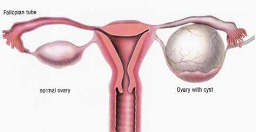
Ovarian cysts are small fluid-filled sacs that develop in a woman's ovaries. Most cysts are harmless, but some may cause problems such as rupturing, bleeding, or pain; and surgery may be required to remove the cyst(s). It is important to understand the function of the ovaries and how these cysts may form.

Women normally have two ovaries that store and release eggs. Each ovary is about the size of a walnut, and one ovary is located on each side of the uterus. One ovary produces one egg each month, and this process starts a woman's monthly menstrual cycle. The egg is enclosed in a sac called a follicle. An egg grows inside the ovary until estrogen (a hormone), signals the uterus to prepare itself for the egg. In turn, the lining of the uterus begins to thicken and prepare for implantation of a fertilized egg resulting in pregnancy. This cycle occurs each month and usually ends when the egg is not fertilized. All contents of the uterus are then expelled if the egg is not fertilized. This is called a menstrual period.

In an ultrasound image, ovarian cysts resemble bubbles. The cyst contains only fluid and is surrounded by a very thin wall. This kind of cyst is also called a functional cyst, or simple cyst. If a follicle fails to rupture and release the egg, the fluid remains and can form a cyst in the ovary. This usually affects one of the ovaries. Small cysts (smaller than one-half inch) may be present in a normal ovary while follicles are being formed.

Ovarian cysts affect women of all ages. The vast majority of ovarian cysts are considered functional (or physiologic). This means they occur normally and are not part of a disease process. Most ovarian cysts are benign, meaning they are not cancerous, and many disappear on their own in a matter of weeks without treatment. While cysts may be found in ovarian cancer, ovarian cysts typically represent a harmless (benign) condition or a normal process. Ovarian cysts occur most often during a woman's childbearing years.

The most common types of ovarian cysts are the following:

Follicular cyst: This type of simple cyst can form when ovulation does not occur or when a mature follicle involutes (collapses on itself). A follicular cyst usually forms at the time of ovulation and can grow to about 2.3 inches in diameter. The rupture of this type of cyst can create sharp severe pain on the side of the ovary on which the cyst appears. This sharp pain (sometimes called mittelschmerz) occurs in the middle of the menstrual cycle, during ovulation. About one-fourth of women with this type of cyst experience pain. Usually, these cysts produce no symptoms and disappear by themselves within a few months.


Corpus luteum cyst: This type of functional ovarian cyst occurs after an egg has been released from a follicle. After this happens, the follicle becomes what is known as a corpus luteum. If a pregnancy doesn't occur, the corpus luteum usually breaks down and disappears. It may, however, fill with fluid or blood and persist on the ovary. Usually, this cyst is found on only one side and produces no symptoms.


Hemorrhagic cyst: This type of functional cyst occurs when bleeding occurs within a cyst. Symptoms such as abdominal pain on one side of the body may be present with this type of cyst.


Dermoid cyst: This is a type of benign tumor sometimes referred to as mature cystic teratoma. It is an abnormal cyst that usually affects younger women and may grow to 6 inches in diameter. A dermoid cyst can contain other types of growths of body tissues such as fat and occasionally bone, hair, and cartilage. These cysts can become inflamed. They can also twist around (a condition known as ovarian torsion), compromising their blood supply and causing severe abdominal pain.


Endometriomas or endometrioid cysts
: Part of the condition known as endometriosis, this type of cyst is formed when endometrial tissue (the lining tissue of the uterus) is present on the ovaries. It affects women during the reproductive years and may cause chronic pelvic pain associated with menstruation.

-Endometriosis is the presence of endometrial glands and tissue outside the uterus.
-Women with endometriosis may have problems with fertility.
-Endometrioid cysts, often filled with dark, reddish-brown blood, may range in size from 0.75-8 inches.


Polycystic-appearing ovary: Polycystic-appearing ovary is diagnosed based on its enlarged size - usually twice that of normal - with small cysts present around the outside of the ovary. This condition can be found in healthy women and in women with hormonal (endocrine) disorders. An ultrasound is used to view the ovary in diagnosing this condition.


Polycystic-appearing ovary is different from the polycystic ovarian syndrome (PCOS), which includes other symptoms and physiological abnormalities in addition to the presence of ovarian cysts. Polycystic ovarian syndrome involves metabolic and cardiovascular risks linked to insulin resistance. These risks include increased glucose tolerance, type 2 diabetes, and high blood pressure.

-Polycystic ovarian syndrome is associated with infertility, abnormal bleeding, increased incidences of miscarriage, and pregnancy-related complications.

-Polycystic ovarian syndrome is extremely common and is thought to occur in 4%-7% of women of reproductive age and is associated with an increased risk for endometrial cancer.

Cystadenoma: A cystadenoma is a type of benign tumor that develops from ovarian tissue. They may be filled with a mucous-type fluid material. Cystadenomas can become very large and may measure 12 inches or more in diameter

Look out for causes and treatment of cysts in the next blog post :)

Thursday, July 15, 2010

Focus: Ghana - Afrox (Africa-Oxford) Cancer Foundation


Cancer already causes more deaths each year worldwide than HIV/AIDS, TB and malaria combined. By 2020 there are expected to be 16 million new cases of cancer every year, 70 per cent of which will be in developing countries, which are least prepared to address the growing cancer burden. African countries are the least able of all developing countries to cope, having few cancer care services. Lack of resources and basic infrastructure mean that most Africans have no access to cancer screening

AfrOx's mission is to partner with African countries and assist them with implementing comprehensive cancer prevention and control programmes. AfrOx believes that by forging alliances of individuals, NGOs and governments to share expertise, technology, training and a degree of philanthropic support, much could be done to help reduce global disparities in cancer care.

AfrOx is currently working in Ghana. The four most common cancers in Ghana are liver, prostate, breast and cervical. At present, Ghana has only two oncology centres, one in the capital Accra and one in Kumasi. There are only 4 oncologists for a population of 22 million and no specialist cancer nurses. With your support, we hope to continue our programmes in the following areas:

- Support for national cancer care planning.
- Provision of training and education for healthcare staff.
- Improvement in palliative care.
- The early diagnosis and treatment of cancer.
- Raising public awareness of the early signs of cancer and methods of cancer prevention.

To learn more about Afrox, to donate, or to read Hajiya Turai's speech commending their effort then visit www.afrox.org

Monday, July 12, 2010

Male Cancer : Introduction


Men and Women have different kinds of risks as far as cancer is concerned and this is due to differences in biological features. below is an introduction to types of cancers that could affect Men. Each type would be elaborated on in future posts.

Prostate Cancer:

Prostate belongs to the reproductive system of the males and it is a gland of walnut size. It is located under the bladder, surrounding the urethra.
Age is an important factor in this kind of cancer. As you get older, you are likely to get affected and if you have a family history, it becomes more risky.

Diagnosis is through a rectal test and a blood test which measures antigens which are specific to prostate, and a protein which signals if there is high level of prostate cancer.

This condition is treated through radiation, surgery and hormone therapies.

Lung Cancer:

This can be a severe form of cancer which affects both the genders. Smoking and inhaling second hand smoke are main causes of cancer. Similarly, pollution or asbestos dust can cause the disease. It is diagnosed through X-rays and CAT scans(Computerised Axial Tomography). Surgery can be opted if cancer has not spread.

Colorectal Cancer:

This is cancer of the digestive parts specifically the rectum and colon . It's prone to attack people with family history and genetics. A high fat diet is also said to be a contributing reason.

It is diagnosed through Colonoscopy, which guides a lighted tube with a camera, inserted into the rectum so that large intestines can be examined. The cancer can be treated through colonoscopy, but if it is advanced it may necessitate a portion of colon to be cut out.

Testicular Cancer:

Testicles are a pair of sex glands in males, just under the penis. They produce and store sperm and are the main source of testosteron. This cancer affects men who have un-descended testes or some birth defects.

The cancer can be felt through unusual lumps, enlargement of testicles or pain in that part. Blood test and biopsy are used to confirm this disease. The affected portion can be removed surgically, after which radiation and chemotherapy can follow.

Penile Cancer:

The penis can also be affected by cancer. Human papillomavirus (HPV) infection is linked to penile cancer. Similarly, if you don’t maintain personal hygiene especially in case of uncircumcised men, it can be a risk.

It is diagnosed through physical examination and biopsy tests. Treatment is usually by surgery, chemotherapy, as well as radiation therapy. If surgery is done precisely, the tumor can be removed leaving the penis intact.


Breast Cancer:


Men are at risk of breast cancer just as women are although its very rare. it accounts for less than 1% of all cancers in men, and less than 1% of all diagnosed breast cancers. Most men are between 60 and 70 years of age when they are diagnosed, but cancer may develop at any age. Worldwide, male breast cancer appears to be becoming more common, particularly in younger males. risk factors includes obesity, increased oestrogen, family history, and Gynaecomastia (enlargement of the male breast tissue).

diagnosis is the same as for women and this includes mammography, biopsy, full blood count and CAT scan.

Treatment is also similar to that in females - by radiotherapy, chemotherapy and hormonal therapies.

Thursday, June 10, 2010

Detecting Breast Cancer


Breast cancer is one of the major causes of death in african women today. early detection is one of the reasons why some cancer victims have a greater chance of survival.Its also been found that self-examination increases the survival rate of breast cancer.

below are some tips on how to do a self-examination of your breast to spot the first signs and symptoms of breast cancer. What you are trying to lookout for are lumps, knots, thickening or swellings which indicate a growing cancer cell inside the breast.

Tip #1:

Lie down on your back and put a pillow under your left shoulder. This will put you in an elevated position after. Raise and place your left hand under your head. Your left breast should be stretched and flattened a bit in this position. Using your right hand, examine your left breast for lumps in a circular motion from the rim of your breast area to the nipple, including the armpit area. Repeat this procedure by reversing the position to the right side of your breasts.

Tip #2:

Examine your breasts by facing a mirror. You can either leave your hands relaxed on the sides or raise them up. Look closely at your breasts for any changes such as the texture, color and orientation. In addition, observe if there is any skin irritation, retraction of nipples, dimpling or bulge and swelling or soreness.

Tip #3:

Examine your breast by contracting your chest muscles. Raise your arm near your forehead and clasp both hands. Then squeeze palms tightly together to make the muscles contract. You must do this while facing the mirror to see if there are any bulges. Another way is to place your palms on the sides of your hips and press them downward. As the muscles contract, check again for bulges.

Tip #4:

Periodically examine your breasts while taking a shower. Raise your one hand overhead, while your other hand carries out examination in a circular motion. Carryout this procedure on both breasts. Why do this in the shower? Because with soap applied on your skin, it can help your hand glide smoothly and feel the lump easily


Reference: cancer reasearch UK